


Prostate
The prostate is a part of the male reproductive system. It is located just in front of the rectum, below the bladder, and surrounds the urethra. The two seminal vesicles, one left and one right, open into the back of the prostate.
With increasing age the prostate can become enlarged (benign prostatic hyperplasia) and cause pain on passing water.
Prostate carcinomas
Prostate carcinomas occur mostly at the edge of the gland tissue and thus, unlike benign prostatic hyperplasia, cause hardly any pain. Complications only occur when the disease advances, as a result of the tumour spreading, and in this case the pain mostly arises from metastases.
Prostate cancer can only be cured if metastasis formation has not yet taken place. It’s therefore very important to identify the disease in good time. The PSA test, a blood test, is available as an early detection examination.
Diagnosis
The most important examination is feeling the prostrate through the rectum. The tumour can then be detected as a hardening of the gland.
If the doctor discovers any suspicious change, or if repeated increase of PSA arouses strong suspicions of tumour disease, a histological examination is necessary. Here, samples are taken from the prostate during an outpatient procedure.
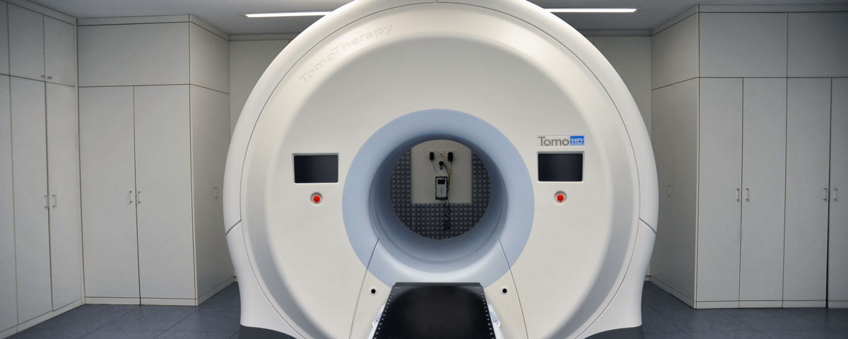
If a cancer of the prostate is detected, it is necessary to know the precise extent of the disease in order to carry out treatment as safely and, at the same time, as harmlessly as possible. PET/CT is the only procedure that can reliably reveal not only tumours in the prostate, but also lymph nodes and other remote metastases. The
PSMA ligand (Prostate Specific Membrane Antigen), which is labelled with gallium 68 and is slightly radioactive, is used for this examination. Its distribution throughout the entire body is measured using PET. This means tumour disease can also be detected earlier and considerably more accurately using PET/CT than using CT alone, which does not show metabolic processes, only structural changes. However, by combining CT with PET, a noticeable accumulation of labelled PSMA ligands from the PET can be accurately matched to the anatomical structures shown in the CT image. The combination of PET and MRT can also be used here. Both hybrid imaging methods are used at the DTZ in Berlin-Friedrichshain.
 Practical guide: The DTZ at a glance


